Yu-li Cai1 ![]() ,
Xing-cai Zhang2
,
Xing-cai Zhang2
For correspondence:- Yu-li Cai Email: caiyuli133494@163.com Tel:+8653168616666
Received: 21 July 2015 Accepted: 11 June 2016 Published: 31 July 2016
Citation: Cai Y, Zhang X. Protective effect of Rhizoma drynariae extract on osteoporosis in ovariectomized rat model. Trop J Pharm Res 2016; 15(7):1447-1452 doi: 10.4314/tjpr.v15i7.13
© 2016 The authors.
This is an Open Access article that uses a funding model which does not charge readers or their institutions for access and distributed under the terms of the Creative Commons Attribution License (http://creativecommons.org/licenses/by/4.0) and the Budapest Open Access Initiative (http://www.budapestopenaccessinitiative.org/read), which permit unrestricted use, distribution, and reproduction in any medium, provided the original work is properly credited..
Purpose: To investigate the therapeutic effect of Rhizoma Drynariae extract (RDE) on ovariectomy-induced osteoporosis in rats.
Methods: Female Sprague-Dawley rats were randomly assigned to a sham-operated group (control) and five ovariectomy (OVX) subgroups: OVX with vehicle (OVX), OVX with 17ß-estradiol (E2, 25 µg/kg/day), and OVX with RDE doses (40, 80, and 160 mg/kg/day). Daily oral administration of E2 or RDE started 4 weeks after OVX and lasted for 16 weeks. The bone mineral density (BMD) of the L4 vertebrae and right femurs was estimated. The length of each femur was measured with a micrometer gauge, and the center of the diaphysis determined. Three representatives L4 vertebrae were selected to evaluate the trabecular microarchitecture. Serum alkaline phosphatase (ALP), urinary calcium (U-Ca), urinary phosphorus (U-P), urinary creatinine (Cr) and osteocalcin (OC) levels were measured.
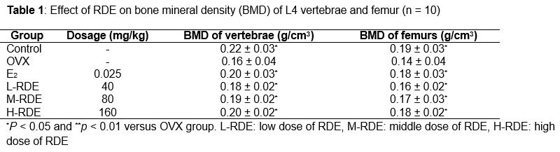
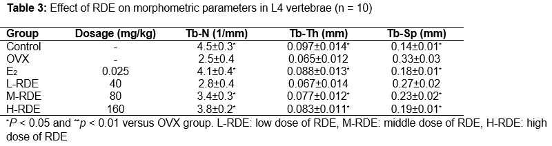
Results: The study showed that high-dose of RDE significantly inhibited the bone mineral density (BMD) reduction of L4 vertebrae (0.20 ± 0.02 g/cm3, p < 0.05) and femurs (0.18 ± 0.02 g/cm3, p < 0.05) caused by OVX and prevented the deterioration of trabecular microarchitecture (p < 0.05), which were accompanied by a significant decrease in skeletal remodeling (p < 0.05) as evidenced by the lower levels of bone turnover markers. High-dose of RDE improved morphometric parameters, namely, Tb-N (3.8 ± 0.2 mm, p < 0.05), Tb-Th (0.083 ± 0.011 mm, p < 0.05) and Tb-Sp (0.19 ± 0.01 mm, p < 0.05) in L4 vertebrae significantly. The present study indicates that the administration of RDE at higher doses over a 16-week period can prevent OVX-induced osteoporosis in rats without hyperplastic effects on the uterus.
Conclusion: Thus, RDE is a potential natural alternative for postmenopausal osteoporosis treatment in elderly women.
Introduction
Osteoporosis is a systemic skeletal disease characterized by reduced bone mass and micro-architectural deterioration of bone tissue with a consequent increase in bone fragility and susceptibility to fractures [1]. According to data released by the World Health Organization (WHO), osteoporosis affects approximately million people throughout Europe, the USA, and Japan [2]. The incidence of osteoporosis increases dramatically with life expectancy. Accordingly, the risk of osteoporotic fractures and their associated costs is rising rapidly due to population aging [3]. In the elderly, hip fractures are closely associated with mortality [4]. Hormone deficiency is known to impair cancellous metaphyseal bone and reduce BMD in humans and animals; therefore, estrogen deficiency in post-menopausal women has been regarded as a critical cause of this population’s susceptibility to osteoporosis [5]. Osteoporosis is twice as common in women as in men [6], and approximately one in three women over 50 years old experiences an osteoporotic fracture in her life time [7].
Clinically, hormone replacement therapy (HRT) has been a popular therapeutic strategy designed for postmenopausal osteoporosis [8,9]. However, the long-term application of HRT has potential malignant effects on reproductive tissues [10-13]. Other medicines that stimulate bone formation (e.g., growth hormone, sodium uoride, and parathyroid hormone) or inhibit bone resorption (e.g., bisphosphonates and calcitonin) may prevent bone loss progression in established osteoporosis. However, these medications are not effective for a large proportion of the world population, especially in developing countries, and these drugs have side effects, such as gastrointestinal reactions, cancers, osteonecrosis of the jaw, and reduced skeletal strength [14,15]. Consequently, to substitute or reduce the medicines used currently, there are efforts to develop new drugs with improved therapeutic efficacy, fewer undesirable side effects, and lower price.
R. drynariae, the dried rhizome of Drynaria fortune (Kunze) J.Sm. (Gu-Sui-Bu in Chinese), has been widely used as a kidney-tonifying and anti-osteoporosis herb for the treatment of nephrasthenia syndrome [16], osteoporosis [17,18] and bone fracture [19] for thousands of years in China. Therefore, the aim of the present study was to systematically evaluate the effect of RDE on osteoporosis induced by OVX in rats.
Methods
Preparation of Rhizoma Drynariae extract
The herbal samples of Rhizoma Drynariae were collected from Bozhou City, Anhui Province in China in July 2014. Taxonomic identification of the plant was performed by Professor Kang Hu of Shandong University of Traditional Chinese Medicine, in China. A voucher specimen (no. RDE 201409015) was deposited in Shandong University of Traditional Chinese Medicine, China for future reference.
One batch of herbal samples Rhizoma drynariae was dried in an oven. Aqueous extract of RDE was obtained by steeping the dried Rhizoma drynariae in water at 60 oC three times for 1 h each. Then drying the extracted fluid in an oven and freeze-drying the last extract. The same batch was extracted for 3 times. One gram powder was equivalent to about 1.5 g crude samples. The yield was 66.67 %.
Animals and treatment
Healthy three-month-old female Sprague-Dawley rats (weighing 220 ± 10 g).Were provided by the Experimental Animal Center of Shandong Province (Certificate no. SYXK 2004-0003). The animals had free access to feed and water, and were allowed to acclimatize for at least one week before use. The rat experiment was approved by the Animal Care and Use Committee of Shandong University of Traditional Chinese Medicine (approval ref no. 20120804) and was carried out in compliance with the Directive 2010/63/EU on the handling of animals used for scientific purposes [20].
60 rats were randomly divided into six groups of ten individuals: a sham-operated group (control) and five ovariectomy (OVX) subgroups, that is, OVX with vehicle (OVX), OVX with 17ß-estradiol (E2, 25 mg/kg/day), and OVX with RDE doses (40, 80 and 160 mg/kg/day). Daily oral administration of E2 or RDE started 4 weeks after OVX and lasted for 16 weeks.
Bone mineral density measurement
The BMD of the L4 vertebrae and right femurs was estimated using dual-energy x-ray absorptiometry scanning (DEXA, GE Healthcare, USA) with small animal measurement. The measurements were expressed as grams of mineral contents per cm2 of surface area. Scans were performed by the same blinded technician.
Three-point bending test
Before mechanical testing, the rats were sacrificed by cervical spondylosis. Then the left femurs were slowly thawed at room temperature. The length of each femur (distance from the intermalleolar to the intercondylar region) was measured with a micrometer, and the center of the diaphysis was determined.
Micro-CT analysis
Based on the BMD, three representatives L4 vertebrae from each group were selected to evaluate the trabecular microarchitecture using eXplore Locus SP preclinical specimen microcomputed tomography (MicroCT, GE Healthcare, USA). Before the scans, the bones were positioned with gauze in the sample holder and allowed to reach room temperature. The L4 vertebrae were scanned from the anterior endplate in the posterior endplate direction (22 um/slice). The isotropic voxel resolution of bone was 22 um3. The volume of interest (VOI) was selected as a region 25 slices away from the anterior endplate to the posterior endplate, ranging to 125 slices. The three-dimensional images were reconstructed with the purpose of visualization and display. After analyzing the VOI, morphometric bone parameters, including trabecular number (Tb-N), trabecular separation (Tb-Sp), trabecular thickness (Tb-Th) were obtained. The VOI analysis was performed blindly by the same operator.
Biochemical analysis of serum and urine specimens
The serum of rats were collected by picking eyeball, and the urine of rats were collected by stimulating back. The levels of serum alkaline phosphatase (ALP), urinary calcium (U-Ca), urinary phosphorus (U-P), and urinary creatinine (Cr) were measured on an automatic analyzer (Ciba-Corning 550, USA) using a diagnostic reagent kit. Serum osteocalcin (OC) concentration was determined using a rat OC ELISA kit (SanClemente, CA, USA).
Statistical analysis
The data are expressed as the mean ± SD. Statistical analysis was performed using one-way ANOVA combined with Bonferroni’s multiple comparison test using SPSS 16.0. Differences were considered statistically significant at p < 0.05.
Results
BMD of L4 vertebrae and femur
The BMD of the L4 vertebrae and femurs is presented in . These results demonstrate that OVX significantly decreased the BMD in the L4 vertebrae and femurs compared to the sham group (p < 0.05). Compared to the OVX group, RDE treatment significantly prevented the BMD decrease in OVX-induced L4 vertebrae and femurs (all p < 0.05) in a dose-dependent manner. E2 also significantly increased the BMD of the L4 vertebrae and femurs (both p < 0.05), which was similar to that observed in the H-RDE group (p > 0.05).
Mechanical testing of femur
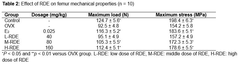
The results of the femur mechanical testing are presented in . Compared with the sham group, 16 weeks of estrogen deficiency significantly decreased the maximum load and maximum stress (both p < 0.05). Meanwhile, higher dosage of RDE treatments (80 or 160 mg/kg/day) markedly prevented the OVX-induced tendency to decrease these parameters (p < 0.05). E2 also increased these biomechanical parameters, which were significantly higher than those of the OVX group (p < 0.05). It is worth noting that the increase in maximum load observed for the H-RDE group was similar to that of E2 (p > 0.05).
Micro-CT data for L4 vertebrae
Analysis of the representative samples () indicated that OVX resulted in the deterioration of the trabecular bone microarchitecture, as demonstrated by the reduced Tb-N and Tb-Th compared with the sham group (both p < 0.05). In contrast, Tb-Sp was significantly increased in response to OVX compared to the sham group (p < 0.05). Higher dosage of RDE treatment (80 or 160 mg/kg/day) significantly improved the microarchitecture deterioration mentioned above (p < 0.05), while E2 also reversed these parameters to the similar degree as that in the H-RDE group (p > 0.05).
Biochemical profile of serum and urine specimens
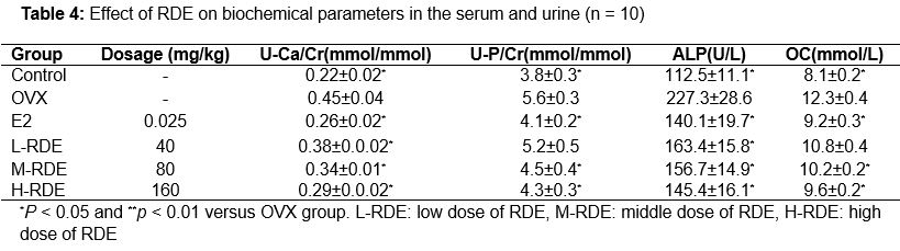
Data presented in show the effect of RDE on biochemical parameters in the serum and urine of OVX rats. Serum U-Ca/Cr, U-P/Cr, ALP, and OC levels were significantly increased in the OVX group compared to the sham group (p < 0.05). All three RDE doses significantly decreased the U-Ca/Cr and ALP levels (p < 0.05) in a dose-dependent manner. Higher dosage of RDE treatment (80 or 160 mg/kg/day) decreased the U-P/Cr and OC levels (p < 0.05). Again, E2 administration also reversed these increases which were statistically significant. It is worth noting that the reductions in U-P/Cr, ALP, and OC in the H-RDE group were similar to those observed for E2 (all p > 0.05).
Discussion
The high incidence, serious complications, financial burden, and dramatically decreased living quality indicate the severity of osteoporosis in humans. Despite the pharmacological and clinical advantages of HRT as a widely accepted therapeutic strategy for osteoporosis, serious side effects of long-term application have also been reported. Therefore, the development of new preventive and therapeutic drugs for osteoporosis is urgently needed. In recent decades, Chinese medicinal herbal extracts have been extensively investigated for their pharmacological effects related to bone protection. Bone remodeling is the biologic process that mediates changes in the traits that influence bone strength [1]. Any interruption in bone remodeling, such as menopause, will disturb the balance between formation and resorption and cause bone mass loss [21,22]. Therefore, we used OVX rats as an animal model for human osteoporosis in vivo experiments. It has been reported that statistically significant bone loss can be seen after 30 days [23], so treatment was initiated 4 weeks after OVX. Consistent with other studies, OVX caused significantly higher body weights in our present study, which may be attributed to fat deposition caused by the lack of estrogen [24]. Previous studies suggest that estrogen plays an important role in stimulating the differentiation of progenitor cells through the osteoblast lineage but not the adipocyte lineage [25].
Decreased BMD is one of the major factors jeopardizing bone strength, resulting in increased susceptibility to fractures [26]. Thus, BMD measurement can best predict fracture risk [27]. The results in the present study showed that OVX reduced BMD in the right femurs and L4 vertebrae, which are rich in trabecular bone, while treatment with RDE dose-dependently prevented the decreases in BMD. Although BMD is among the strongest predictors of facture resistance, both empirical observations and theoretical analyses show that the biomechanical properties of bone and trabecular microarchitecture influence trabecular bone strength as well [28-30]. Three-point bending tests of the left femurs in our study indicated that the higher croc in doses (80 or 160 mg/kg/day) prevented the OVX-induced tendency toward decreased biomechanical parameters.
Moreover, measurements of structural parameters using micro-CT also showed that treatment with RDE effectively restored the trabecular micro architectural properties compared to the OVX group. In addition, measurement of bone markers plays a role in osteoporosis diagnosis and treatment [31]. Bone mass loss, as evidenced by enhanced levels of ALP, OC, U-Ca/Cr, and U-P/Cr, indicated upregulation of bone turnover by OVX. The bone turnover markers above were dose-dependently reversed by RDE, indicating a reduction in bone turnover rate after treatment of RDE. Our results suggested RDE’s effectiveness in treating osteoporosis in menopausal women.
Conclusion
This study demonstrate that daily oral administration of RDE over a 12-week period prevents estrogen deficiency-induced bone loss, inhibits the deterioration of trabecular microarchitecture, maintains the biomechanical competence of bone, decreases bone turnover rate, and does not stimulate an unwanted proliferation of the uterine tissues. Our findings indicate that RDE has the potential to be further developed as a natural alternative for postmenopausal osteoporosis management in elderly women.
References
Archives


News Updates